
인류의 건강과 생명공학 발전을 위해 봉사하는 기업 드림셀
We Serving the Health and Biotechnology of Humanity
We Serving the Health and Biotechnology of Humanity
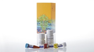
제품코드 : J3160
제품정보 : Energy Metabolism Assay
The Triglyceride-Glo™ Assay is a bioluminescent assay for rapid and sensitive measurement of triglycerides in cultured cell lysates and other biological samples, such as cell culture medium, serum and tissue homogenates. The assay is ideal for measuring triglyceride accumulation and clearance in normal and pathological conditions. Examples include adipocytes, liver samples or cell culture liver models where excess triglyceride accumulation causes steatosis, an early hallmark of nonalcoholic fatty liver disease (NAFLD) and nonalcoholic steatohepatitis (NASH).
The Triglyceride-Glo™ Assay detects triglyceride levels by measuring glycerol that is released from an enzymatic reaction with a lipase: one mole of glycerol per mole of triglyceride. Glycerol is measured in a coupled reaction scheme that links the production of NADH to the activation of a proluciferin that produces light with luciferase. The amount of triglyceride is determined from the difference of glycerol measured in the absence (free glycerol) and presence (total glycerol) of lipase. Lipase converts triglyceride (TAG) to glycerol. Glycerol kinase and glycerol-3-phosphate dehydrogenase are used to generate NADH. In the presence of NADH, Reductase enzymatically reduces a proluciferin Reductase Substrate to luciferin. Luciferin is detected in a luciferase reaction using Ultra-Glo™ Luciferase and ATP, and the amount of light produced is proportional to the amount of glycerol in the sample.

Adipocytes differentiated from 3T3L1-MBX fibroblasts were measured with or without an organic extraction method and the Triglyceride-Glo™ Assay. One set of wells was extracted with methanol, transferred to a glass vial and allowed to evaporate to dryness overnight.
No organic extraction was performed on the other set of wells. Both samples were then extracted with the Lysis Solution and triglyceride levels were measured. The triglyceride recovery without organic extraction was higher and less variable over six replicates.


Quantifying triglyceride levels in liver cells is used in liver disorder research for disorders such as non-alcoholic fatty liver disease (NAFLD) and non-alcoholic steatohepatitis (NASH). Here, human liver microtissues are used to model steatosis of liver cells and quantitating triglyceride levels.
3D InSight™ human Liver Microtissues (MT, InSphero, ~1150 cells/well) were incubated for 3 and 10 days in serum-free medium containing either physiological (LG/LI) or supraphysiological (LG/HI) levels of glucose and insulin and supplementation with either free fatty acids bound to BSA (FFA) or low density lipoprotein plasma fraction (LDL). The microtissues were washed twice in PBS and assayed for total glycerol content according to the standard protocol, as intracellular glycerol content does not contribute significantly to the total glycerol content. Values were plotted as concentration of triglyceride per MT. The data was provided by InSphero, AG, Zurich, Switzerland.
The lipid accumulation in differentiating adipocytes was monitored with Oil Red O staining and the Triglyceride-Glo™ Assay. Parallel plates of 3T3L1-MBX fibroblasts were differentiated following standard protocol.
At days 5, 12, 14 and 21 (Panels A–D, respectively) the parallel plates were either stained with Oil Red O or assayed with the Triglyceride-Glo™ Assay for total glycerol content. The Triglyceride-Glo™ Assay quantitatively measures triglycerides levels in differentiated adipocytes.
The simple sample preparation protocol includes only a detergent lysis step and sample dilution prior to triglyceride quantification. No organic extraction or extreme heating steps are required.

Triglyceride-Glo™ Assay Technical Manual, TM600
Triglyceride-Glo™ Assay Quick Protocol FB229