
인류의 건강과 생명공학 발전을 위해 봉사하는 기업 드림셀
We Serving the Health and Biotechnology of Humanity
We Serving the Health and Biotechnology of Humanity
제품코드 : W6040
제품정보 : Inflammation Assay
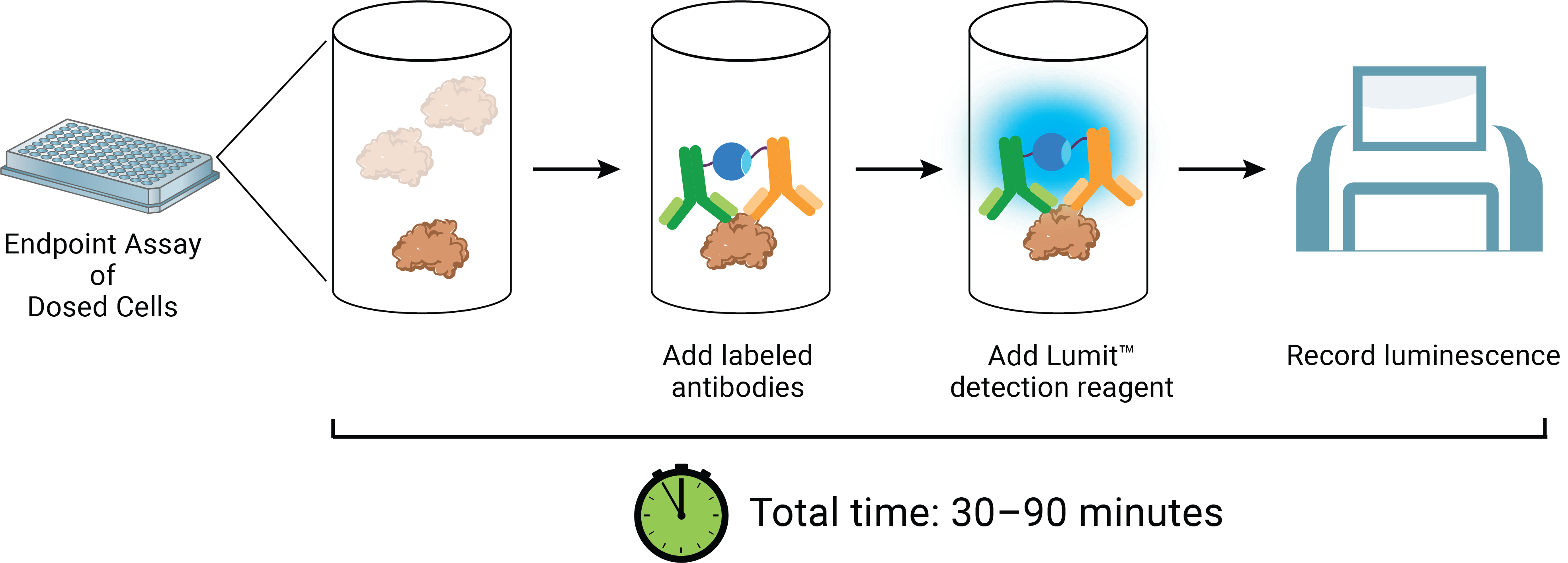
The Lumit™ IFN-γ Immunoassay quantitatively measures released IFN-γ in cell culture samples using a simple, no-wash protocol. Just add labeled antibodies to the sample, add detection reagent and read luminescent signal using a standard plate-reading luminometer. The entire protocol is completed in less than 70 minutes! The assay can be used directly on cells in culture or culture medium transferred to a separate assay plate.
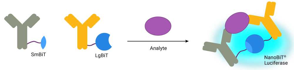
Primary antibodies to IFN-γ were selected for their specific and sensitive detection and labeled with the LgBiT and SmBiT subunits of NanoBiT® Luciferase. In the presence of IFN-γ, the subunits are brought together to form an active luciferase enzyme. Addition of optimized substrate generates a bright luminescent signal proportional to IFN-γ levels.
The pg/ml to ng/ml linear range of Lumit™ detection covers expected concentrations for most cell models.

Assay | IFN-γ Human Immunoassay | |
Limit of Detection (LOD) | 1.7pg/ml (3 SD above background) | |
Dynamic Range | 7.3pg/ml – 10ng/ml | |
Minimal Detectable Dose (MDD) | 1.1pg/ml (2 SD above background) | |
Assay Time | 70 minutes | |
Sample Type | Cell culture supernatants | |
SD=Standard Deviation

Purified CD8+ T cells (effector cells) were combined with Raji B cells (target cells) and a serial dilution of Blincyto® (CD3 and CD19 bispecific T cell engager). The Lumit™ IFN-γ (Human) Immunoassay reagents were added directly to treated cell wells to detect IFN-γ release from the T cells.

Human PBMCs were plated in a 96-well plate in 100µl and treated for 24 hours with cell stimulation cocktail (CSC), lipopolysaccharide (LPS) or R848. Four 20μl aliquots were transferred to a 384-well plate for detection of IL-2, IL-6, IFN-γ and TNF-α with the corresponding Lumit™ Cytokine Immunoassay. This simple approach for profiling cytokine release can be applied to multiple cell models.
We offer a growing portfolio of assays to detect cytokines in cell culture samples.
Can’t find your target in the list?
Please enquire about early access to new assays in development.
For a biotech that understands time is of significant value, the Lumit™ Immunoassays are an essential way for us to gather reliable data fast so that we can reach conclusions and make decisions sooner.
Jonathan Chow, Principal Scientist, Corner Therapeutics