본문 바로가기 주메뉴 바로가기

인류의 건강과 생명공학 발전을 위해 봉사하는 기업 드림셀

We Serving the Health and Biotechnology of Humanity
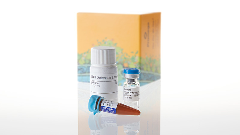
제품정보

> 제품정보 > 시약 > 국내 공식 대리점 > LDH-Glo™ Cytotoxicity Assay, 10ml
제품명

LDH-Glo™ Cytotoxicity Assay, 10ml

제품코드 : J2380

제품정보 : Cytotoxicity Assay

  • 발광 기반의 LDH assay로 원하는 시간에 적은 양의 배지를 sampling하여 시간에 따른 세포 독성변화를 확인할 수 있습니다.
  • [J2380] 10ml, [J2381] 50ml

제품문의

  • 전화번호
    2292-8870

제품상세

Simple, More Sensitive LDH Assay Method to Determine Cytotoxicity

  • Detects LDH release from small numbers of cells, including 3D cell models
  • Monitors cytotoxicity from the same sample over time
  • Provides more data per well through multiplexing with other cell-based assays

Monitor Cytotoxicity from Small Numbers of Cells

The LDH-Glo™ Cytotoxicity Assay is a bioluminescent plate-based assay for quantifying lactate dehydrogenase (LDH) release into the culture medium upon plasma membrane damage. The bioluminescent detection is more sensitive than colorimetric or fluorescent methods, allowing accurate detection of LDH from a small number of cells, including primary cells and 3D cell cultures.

This LDH assay involves removing only a small amount of cell media (2–5µl) from each treated well, allowing you to get more data by sampling the same well over time, and by using the remaining media and cells for other cell-based assays.

How the Assay Works

LDH released from damaged cells catalyzes the oxidation of lactate with concomitant reduction of NAD+ to NADH. Reductase uses NADH and reductase substrate to generate luciferin, which is converted to a bioluminescent signal by Ultra-Glo™ rLuciferase. The luminescent signal generated is proportional to the amount of LDH present.
Lactate dehydrogenase detection with LDH-Glo cytotoxicity assay 15079ma-w

"The Promega LDH-Glo™ Assay is a reliable and easy-to-follow kit to use when measuring cell death. We use this in our lab and, compared to others, it really helps us get reliable data. Overall, we would highly recommend this kit."


LDH-Glo Customer

Monitor Cytotoxicity in 3D Cell Cultures

The LDH-Glo™ Cytotoxicity Assay is well-suited for measuring LDH released from small numbers of membrane-damaged cells. Here is an example of measuring drug-induced toxicity in HCT116 spheroids. The time-dependent toxicity measurements were performed by repeatedly sampling media from the same wells.
Measuring drug-induced toxicity in HCT116 spheroids using the bioluminescent ldh assay 15086mb-wb
HCT116 spheroids were formed using 2,500 cells/well in Corning 384-well Ultra-Low Attachment plates and treated with Doxorubicin. 2.5µl samples were collected from the same wells at the indicated time points and frozen in LDH Storage Buffer at a 1:10 dilution. Samples were thawed and further diluted 2.5-fold in LDH Storage Buffer before measuring LDH levels.
See more data: Learn how this assay is used with different model systems, including SKBR3 cells to assess an antibody drug conjugate and in 3D human liver microtissues to monitor cytotoxicity over time, in the poster: New Sensitive Luminescent LDH-release Assay that Enables “Real-Time” Sampling from Individual 3D Microtissues, presented at SLAS Europe 2018.

Detect Target Cell Cytotoxicity in ADCC Assays

The LDH-Glo™ Cytotoxicity assay can be used to assess cell death due to many mechanisms of action of biologics treatments, including complement dependent cytotoxicity (CDC), antibody-dependent cell-mediated cytotoxicity (ADCC) and antibody-drug conjugate cytotoxicity. In this example, the LDH-Glo™ Assay was used to detect ADCC killing of target cells using the biologic rituximab.
 
Detection of target cell cytotoxicity in ADCC assays using a bioluminescent ldh assay method 15087mb-wa
Cells were plated at a 20:1 Effector (PBMC):Target (Daudi) ratio and treated with varying concentrations of rituximab (RTX) in triplicate for 4 or 6 hours. Medium samples (2.5µl) from the same wells were removed and diluted into LDH Storage Buffer (47.5µl) and frozen. 25µl of thawed samples were added to an equal volume of LDH Detection Reagent and luminescence was recorded after 60 minutes incubation at room temperature. Concentration of rituximab versus RLU was plotted and data was fit to 4PL curve to calculate EC50
Detection of target cell cytotoxicity in ADCC assays using a bioluminescent ldh assay method 15087mb-wb
RLU values from control wells (performed on the same plate as the rituximab treatment) indicate that there is minimal luminescent signal from target or effector cells alone, and that addition of rituximab is required for cell killing. ADCC = Effector cells +Target cells + RTX. RLU = Relative Luminescence Units.


아래 링크를 클릭하시면 제품에 대한 자세한 정보를 확인하실 수 있습니다.