인류의 건강과 생명공학 발전을 위해 봉사하는 기업 드림셀
We Serving the Health and Biotechnology of Humanity
We Serving the Health and Biotechnology of Humanity

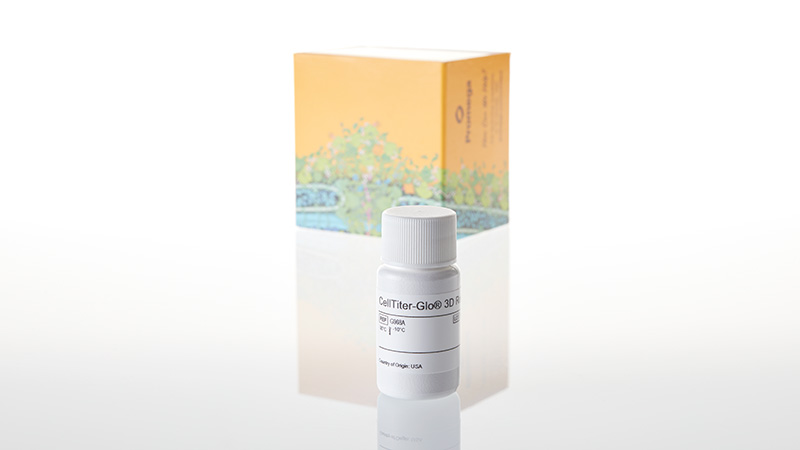
제품코드 : G9681
제품정보 : Cell Viability Assay
The CellTiter-Glo® 3D Cell Viability Assay is designed for determining cell viability in 3D microtissue spheroids. The assay reagent penetrates large spheroids and has increased lytic capacity—allowing more accurate determination of viability compared to other assay methods.
Based on the same reliable chemistry as the classic CellTiter-Glo® Assay, this new 3D assay reagent measures ATP as an indicator of viability and generates a luminescent readout that is much more sensitive than colorimetric or fluorescence-based methods. The simple, 30-minute protocol and ready-to-use reagent allows for fast results.
| Diameter of Spheroid (μm) | Classic CellTiter-Glo® Assay (pmol/microtissue) | CellTiter-Glo® 3D Assay (pmol/microtissue) | Ratio |
|---|---|---|---|
| 188 | 16 ± 4 | 17 ± 4 | 1.10 |
| 386 | 79 ± 3 | 94 ± 11 | 1.19 |
| 459 | 103 ± 2 | 126 ± 11 | 1.22 |
| 565 | 127 ± 3 | 178 ± 17 | 1.40 |
HCT116 colon cancer spheroids were generated by seeding cells in the InSphero GravityPLUS™ 96-well hanging-drop platform and grown for 4 days.
Panel A. An equivalent volume of reagent was added to all samples, and after 5 minutes of shaking, luminescence was recorded at 30 minutes.
Panel B. A 2X concentration of CellTox™ Green Dye was added to CellTiter-Glo® 3D Reagent (left) or ATPlite™ 1Step Reagent (right) prior to sample addition as an indicator of cell lysis and images were acquired at 30 minutes. The spheroids in Panel B are ~300μm in diameter, and the bars in each image represent a distance of 200μm.

Luminescent signals from the CellTiter-Glo® 3D Assay are orders of magnitude above background. Other non-lytic viability assays that measure changes in fluorescence (e.g. alamarBlue®) or absorbance (e.g., MTT) generate signals that are only modestly higher than their no-cell control signals.

HCT116 colon cancer cells seeded into an InSphero GravityPLUS™ 96-well hanging-drop platform and grown to generate ~340μm spheroids.

InSphero Insight™ human liver microtissues (~250μm). All microtissues were assayed using the CellTiter-Glo® 3D, alamarBlue® and MTT assays according to manufacturer's protocols. Total assay times for the CellTiter-Glo® 3D, alamarBlue® and MTT assays were 30 minutes, 3 hours and 8 hours, respectively.
The results of compound screening using the CellTiter-Glo® 3D Assay in hanging-drop, ultra-low attachment plate (ULA) and Matrigel® 3D cultures are shown below. Equivalent results were achieved for all three methods.


HCT116 colon cancer cells were seeded as follows: 400 cells in hanging-drop; 1,000 cells in ULA or Matrigel®. Microtissues were grown for 4 days, treated with compounds for 48 hours, and then assayed with the CellTiter-Glo® 3D Reagent. Luminescence was recorded at 30 minutes.
Four hundred HCT116 colon cancer cells were seeded into each of 60 wells of a 96-well InSphero GravityPLUS™ hanging-drop plate and incubated for 4 days to form 60 spheroids (~350μm in diameter). Half of the spheroids were treated with 100μM panobinostat (black squares), and the other half were treated with vehicle (1% DMSO, orange squares). After 48 hours, all samples were assayed with the CellTiter-Glo® 3D Reagent. The CellTiter-Glo® 3D Assay provided a Z´-factor of 0.81.
